

A Review of Coupled Thermo-Mechanical Behaviors of Brain Tissue
-
摘要:
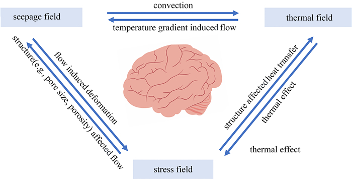
脑组织是由固相与液相组成的饱和含液多孔材料,固、液、生理环境(尤其温度)之间相互作用,具体表现为脑组织内部温度场、渗流场、应力场相互影响的热-力耦合行为,故阐明脑组织的热-力耦合行为是理解大脑功能和疾病病理的关键.该文首先介绍了脑组织的传热学和力学性质,重点关注实验测量及应变率和温度的影响;其次,总结了描述脑组织热-力耦合行为的数理模型,包括力学模型、传热学模型和热-力耦合模型;最后,对该重要学科交叉领域进行了总结和展望.
Abstract:The brain is the highest nerve center regulating physiological behaviors and functions. Brain tissue is a saturated porous material composed mainly of solid phase and liquid phase. Interactions between the solid phase, the liquid phase and the physiological environment (temperature in particular) are manifested in the coupled thermo-mechanical behaviors of brain tissue, and affected by internal temperature, seepage and stress fields. Characterization of the coupled thermo-mechanical behaviors of brain tissue is the key to understanding brain function and disease pathology. Firstly, the thermal and mechanical properties of brain tissue measured via different experimental methods were introduced, with a particular focus placed upon the effects of the strain rate and the temperature. Theoretical and numerical models describing the coupled thermo-mechanical behaviors of brain tissues were then summarized, including mechanical models, heat transfer models and coupled thermo-mechanical models. Finally, this important multidisciplinary field was summarized and prospected.
-
Key words:
- brain tissue /
- mechanical property /
- thermophysical property /
- thermo-mechanical coupling
edited-byedited-by1) (我刊编委卢天健来稿) -


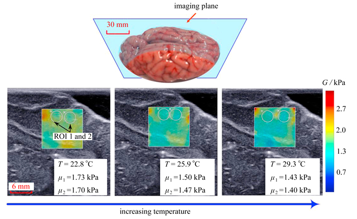
图 4 脑组织剪切模量随温度的变化趋势:图中标注了成像平面上的两个测量区域(ROI 1和ROI 2,白色圆圈)及相应的测量结果μ1和μ2[78]
注 为了解释图中的颜色,读者可以参考本文的电子网页版本,后同.
Figure 4. The shear modulus of brain tissue changing with the temperature: 2 ROIs (the white circles) in the imagin plane and corresponding measurement results μ1 and μ2 illustrated in each image[78]

A1 脑组织力学性质
A1. Mechanical properties of brain tissue
力学性质 样品来源 数值 测试方法 测试条件 参考文献 弹性模量
E/kPa牛脑 0.35 非受限压缩法,应变率0.01 s-1 离体 [72] 白质,牛
灰质,牛1.895±0.592
1.389±0.289压痕法,应变率0.004 s-1 室温,离体 [42] 白质,大鼠
灰质,大鼠0.294±0.074
0.454±0.053扫描力显微镜(SFM)压痕法 室温,离体 [73] 白质,猪
灰质,猪1.787±0.186
1.195±0.157压痕法 室温,离体 [74] 牛脑,非灌注
牛脑,灌注46.8±31.3
106.4±73.9离心实验模拟超重 20±2 ℃,离体 [75] 猪脑 8.12~29.46
10.86~41.05
16.08~60.73拉伸法,应变率30 s-1
拉伸法,应变率60 s-1
拉伸法,应变率90 s-1室温22 ℃,离体 [69] 白质,猪 0.114±0.026
2.947
0.155非受限压缩法,平衡模量
非受限压缩法,应变率2 s-1
非受限压缩法,应变率10-6 s-1室温,离体 [71] 人脑 2.704
0.457非受限压缩法,应变率2 s-1
非受限压缩法,应变率0.001 s-1室温,离体 [71] 剪切模量
G/kPa大鼠脑 0.412~0.453 微压痕法, 应变率1.43 s-1 离体 [76] 大鼠脑 0.398~0.626 压痕法 在体/离体 [43] 猪脑 0.195~0.305 振荡剪切法,应变率1 s-1 37 ℃,离体 [66] 辐射冠,猪 0.6~1.0 压痕法,应变率0.064 s-1 室温,离体 [41] 小脑,小鼠
皮质,小鼠
髓质,小鼠
脑桥,小鼠2.48~3.14
4.83~7.67
3.81~4.32
5.66~6.51微压痕法, 应变率10 s-1 室温22 ℃,离体 [63] 白质,猪
灰质,猪
丘脑,猪
中脑,猪0.925~1.209
0.669~0.816
0.943±0.109
0.955±0.137压痕法 室温,离体 [61] 胼胝体,人
辐射冠,人
基底神经节,人
皮层,人0.33±0.18
0.54±0.21
0.56±0.20
1.06±0.36剪切,准静态加载条件 室温,离体 [77] 小脑,小鼠 2.11±1.26
3.15±1.66
3.71±1.23微压痕法,应变率5 s-1
微压痕法,应变率15 s-1
微压痕法,应变率30 s-122 ℃,离体 [67] 皮质,小鼠 4.06±1.69
6.14±3.03
7.05±3.92微压痕法,应变率5 s-1
微压痕法,应变率15 s-1
微压痕法,应变率30 s-122 ℃,离体 [67] 猪脑 0.311±0.055
0.384±0.038
0.486±0.107
0.546±0.119
0.645±0.071压痕法,应变率0.002 s-1
压痕法,应变率0.008 s-1
压痕法,应变率0.030 s-1
压痕法,应变率0.061 s-1
压痕法,应变率0.152 s-1室温,离体 [68] 猪脑,冷冻保存
猪脑,22 ℃保存
猪脑,37 ℃保存1.043±0.271
0.714±0.210
0.497±0.156简单剪切,应变率30 s-1 22 ℃,离体 [59] 猪脑 1.20
0.84
0.82
0.84
0.92横波弹性成像 25 ℃,离体
37 ℃,离体
42 ℃,离体
45 ℃,离体
48 ℃,离体[78] 切线模量
Et/kPa猪脑,冷冻保存
猪脑,37 ℃保存156.7~2 242.9
500.2~4 959.9SHPB高应变率单轴应力压缩实验,应变率(2 487±72) s-1 37 ℃,离体,10%应变 [60] 存储模量
G′/kPa人脑
猪脑1.182~2.224
1.727~3.757磁共振弹性成像 在体
离体[51] 白质,人
灰质,人
脑干,人0.453~1.903
0.507~1.911
1.270~5.048振荡剪切实验 37 ℃,离体 [79] 白质,人
白质,人
白质,人
白质,人0.866
0.793
0.764
0.754流变实验 22 ℃,离体
27 ℃,离体
32 ℃,离体
37 ℃,离体[80] 损耗模量
G″/kPa人脑
猪脑0.631~1.140
1.233±2.534磁共振弹性成像 在体
离体[51] 白质,人
灰质,人
脑干,人0.082~0.443
0.124~0.456
0.255~1.032振荡剪切实验 37 ℃,离体 [79] 白质,人
白质,人
白质,人
白质,人0.233
0.186
0.164
0.152流变实验 22 ℃,离体
27 ℃,离体
32 ℃,离体
37 ℃,离体[80] Poisson比
ν牛脑 0.35 非受限压缩法,应变率0.01 s-1 离体 [72] 人脑,非排水
人脑,排水0.5
0.496非受限压缩法,应变率0.01 s-1 离体 [81] 牛脑,非灌注
牛脑,灌注0.326±0.198
0.370±0.188离心实验模拟超重 20±2 ℃,离体 [75] 牛脑
0.45~0.47
0.67±0.05非受限压缩 室温(~25 ℃),离体,
应变5%~10%
室温(~25 ℃),离体,应变30%[82]  下载: 导出CSV
A2 脑组织热物性
A2. Thermal properties of brain tissue
热物性 样品来源 数值 测试方法 测试条件 参考文献 密度
ρ/(kg·m-3)白质,马脑
灰质,马脑1 038±1.1
1 039±0.9离体,37 ℃ [99] 脑
灰质
白质1 046
1 050
1 040离体,37 ℃ [100] 比热容
c/(J·kg-1·K-1)白质,人脑 3 590
3 610
3 640
3 690DSC 离体,37 ℃
离体,43 ℃
离体,50 ℃
离体,60 ℃[98] 灰质,人脑 3 590
3 650
3 650
3 700DSC 离体,37 ℃
离体,43 ℃
离体,50 ℃
离体,60 ℃[98] 胶质母细胞瘤 3 630
3 790
3 740
3 640DSC 离体,37 ℃
离体,43 ℃
离体,50 ℃
离体,60 ℃[98] 脑
灰质
白质
小脑3 630±74
3 718±36
3 525±73
3 653DSC 离体,60 ℃ [100] 人脑 4 160 DSC 离体,60 ℃ [101] 热导率
λ/(W·m-1·K-1)牛脑 0.524±0.010
0.553±0.004
0.563±0.005
0.567±0.011
0.560±0.006
0.697±0.034
2.005±0.057双针传感器 离体,22 ℃
离体,33 ℃
离体,41 ℃
离体,52 ℃
离体,66 ℃
离体,83 ℃
离体,97 ℃[88] 脑
灰质
白质
小脑0.51±0.02
0.55±0.03
0.48±0.02
0.51±0.00双针传感器 离体,97 ℃ [100] 人脑 0.49 双针传感器 离体,97 ℃ [101] 白质,人
灰质,人
人脑0.502
0.565
0.528探针法 离体 [97] 热扩散系数
a/(10-6 m2·s-1)牛脑 0.136±0.005
0.145±0.001
0.147±0.001
0.149±0.003
0.158±0.003
0.205±0.015
0.373±0.014双针传感器 离体,22 ℃
离体,33 ℃
离体,41 ℃
离体,52 ℃
离体,66 ℃
离体,83 ℃
离体,97 ℃[88] 白质,人
灰质,人
人脑0.134
0.143
0.138探针法 离体 [97] 体积热容
Cv/(MJ·m-3·K-1)3.86±0.06 离体,22 ℃ 3.83±0.03 离体,33 ℃ 3.83±0.04 离体,41 ℃ 牛脑 3.81±0.06 双针传感器 离体,52 ℃ [88] 3.53±0.08 离体,66 ℃ 3.30±0.19 离体,83 ℃ 4.98±0.20 离体,97 ℃ 热膨胀系数
αT/K-1海马体,大鼠 5.5×10⁻⁴
1.37×10⁻³DIC 离体,30~40 ℃
离体,37~40 ℃[102] 狗脑 5×10⁻⁵ 密度测量技术 离体,25~37 ℃ [103]
下载: 导出CSV
A3 脑组织数理模型
A3. Mathematical models for brain tissue
模型 内容 优点 不足 超弹性模型 适用于脑组织等类橡胶材料,主要包括Neo-Hookean模型、Mooney-Rivlin模型、Ogden超弹性模型等 可以捕捉脑组织时间无关的拉压不对称性、非线性和大变形行为 现象学模型,不能很好地描述脑组织的时间依赖性,无法准确描述含液多孔脑组织的双相性 黏弹性模型 描述脑组织的弹性与黏性,松弛模量采用Prony级数进行描述 可以描述脑组织的时间依赖性,预测脑组织受力后的蠕变与松弛现象 无法捕捉脑组织的拉压不对称特性,无法准确描述含液多孔脑组织的双相性 多孔弹性模型 基于Biot理论,将脑组织看作由弹性固相和无黏液相组成的一种饱和含液多孔材料 可以考虑脑组织内部孔隙流体的影响,描述流体和固体间的相互作用 经典的Biot多孔弹性模型不含尺度,故不能准确描述固-液界面作用,且不能描述脑组织的滞回行为 多孔黏弹性模型 结合多孔弹性与黏弹性,描述多孔材料的黏弹性行为 考虑了固相、液相之间的相互作用对时间和长度尺度的依赖性 脑组织的复杂性使得多孔黏弹性模型的建立和参数确定具有挑战性,参数确定需要更多的实验数据支持 Pennes生物传热模型 用于描述生物组织中热传递过程的数学模型 考虑了代谢、血液灌注对传热的影响,简单、有效,具有普适性 在某些情况下过于简化,例如忽略了血管之间的相互作用、组织的各向异性以及血液流动的复杂性 热-力耦合模型 考虑温度、饱和度、孔隙率、微观结构等因素的影响,涉及固相/液相与外界环境温度和热量的相互作用 可以描述脑组织流体流动、传热和力学变形耦合行为 存在研究空白,目前的模型大多是基于肝脏、皮肤等生物组织的热-流耦合或热-固耦合模型,缺乏热-流-固耦合模型,模型建立困难
下载: 导出CSV
-
[1] BUDDAY S, OVAERT T C, HOLZAPFEL G A, et al. Fifty shades of brain: a review on the mechanical testing and modeling of brain tissue[J]. Archives of Computational Methods in Engineering, 2019, 27(4): 1187-1230. [2] GORIELY A, GEERS M G D, HOLZAPFEL G A, et al. Mechanics of the brain: perspectives, challenges, and opportunities[J]. Biomechanics and Modeling in Mechanobiology, 2015, 14(5): 931-965. doi: 10.1007/s10237-015-0662-4 [3] PROCÈS A, LUCIANO M, KALUKULA Y, et al. Multiscale mechanobiology in brain physiology and diseases[J]. Frontiers in Cell and Developmental Biology, 2022, 10: 823857. doi: 10.3389/fcell.2022.823857 [4] SQUIRE L R, BERG D, BLOOM F E, et al. Fundamental Neuroscience[M]. San Diego: Academic Press, 2013. [5] LEI Y, HAN H, YUAN F, et al. The brain interstitial system: anatomy, modeling, in vivo measurement, and applications[J]. Progress in Neurobiology, 2017, 157: 230-246. doi: 10.1016/j.pneurobio.2015.12.007 [6] STUART G, SPRUSTON N, HÄUSSER M. Dendrites[M]. New York: Oxford University Press, 2007. [7] SYKOVÁ E, NICHOLSON C. Diffusion in brain extracellular space[J]. Physiological Reviews, 2008, 88(4): 1277-1340. doi: 10.1152/physrev.00027.2007 [8] KYRIACOU S K, MOHAMED A, MILLER K, et al. Brain mechanics for neurosurgery: modeling issues[J]. Biomechanics and Modeling in Mechanobiology, 2002, 1(2): 151-164. doi: 10.1007/s10237-002-0013-0 [9] BHARAT S, TECHAVIPOO U, KISS M Z, et al. Monitoring stiffness changes in lesions after radiofrequency ablation at different temperatures and durations of ablation[J]. Ultrasound in Medicine & Biology, 2005, 31(3): 415-422. [10] MOHAMMADI A, BIANCHI L, KORGANBAYEV S, et al. Thermomechanical modeling of laser ablation therapy of tumors: sensitivity analysis and optimization of influential variables[J]. IEEE Transactions on Biomedical Engineering, 2022, 69(1): 302-313. doi: 10.1109/TBME.2021.3092889 [11] SINGH S, MELNIK R. Coupled thermo-electro-mechanical models for thermal ablation of biological tissues and heat relaxation time effects[J]. Physics in Medicine & Biology, 2019, 64(24): 245008. [12] XU F, LU T J. Introduction to Skin Biothermomechanics and Thermal Pain[M]. New York: Springer, 2011. [13] 卢天健, 林敏, 徐峰. 牙齿的热-力-电生理耦合行为[M]. 北京: 科学出版社, 2015.LU Tianjian, LIN Min, XU Feng. Thermo-Mechano-Electrophysiological Coupling Behaviors of Teeth[M]. Beijing: Science Press, 2015. (in Chinese) [14] TIAN J, HUANG G, LIN M, et al. A mechanoelectrical coupling model of neurons under stretching[J]. Journal of the Mechanical Behavior of Biomedical Materials, 2019, 93: 213-221. doi: 10.1016/j.jmbbm.2019.02.007 [15] OCHOA D A, MIRANDA B M, CONGER B C, et al. Lunar eva thermal environment challenges[J]. SAE Transactions, 2006, 115: 492-505. [16] ZU EULENBURG P, VAN OMBERGEN A, TOMILOVSKAYA E S, et al. Reply to Ludwig et al: a potential mechanism for intracranial cerebrospinal fluid accumulation during long-duration spaceflight[J]. Biological Sciences, 2019, 116(41): 20265-20266. [17] VAN OMBERGEN A, JILLINGS S, JEURISSEN B, et al. Brain ventricular volume changes induced by long-duration spaceflight[J]. Proceedings of the National Academy of Sciences of the United States of America, 2019, 116(21): 10531-10536. [18] MACMANUS D B, MURPHY J G, GILCHRIST M D. Mechanical characterisation of brain tissue up to 35% strain at 1, 10, and 100/s using a custom-built micro-indentation apparatus[J]. Journal of the Mechanical Behavior of Biomedical Materials, 2018, 87: 256-266. doi: 10.1016/j.jmbbm.2018.07.025 [19] MOHAJER N, WINTER A, GREGORY T M, et al. Experimental validation of a high-G centrifuge system using an advanced wireless human dummy[C]//Proceedings of the 2022 IEEE International Conference on Systems, Man, and Cybernetics (SMC). Prague, Czech Republic, 2022. [20] WANG L, YIN H, DI Y, et al. Human local and total heat losses in different temperature[J]. Physiology & Behavior, 2016, 157: 270-276. [21] ECKER J R, GESCHWIND D H, KRIEGSTEIN A R, et al. The barin initiative cell census consortium: lessons learned toward generating a comprehensive brain cell atlas[J]. Neuron, 2017, 96(3): 542-557. doi: 10.1016/j.neuron.2017.10.007 [22] CHAGOVETZ A A, JENSEN R L, RECHT L, et al. Preliminary use of differential scanning calorimetry of cerebrospinal fluid for the diagnosis of glioblastoma multiforme[J]. Journal of Neuro-Oncology, 2011, 105(3): 499-506. doi: 10.1007/s11060-011-0630-5 [23] BERTALAN G, BOEHM-STURM P, SCHREYER S, et al. The influence of body temperature on tissue stiffness, blood perfusion, and water diffusion in the mouse brain[J]. Acta Biomater, 2019, 96: 412-420. doi: 10.1016/j.actbio.2019.06.034 [24] BARNES J M, PRZYBYLA L, WEAVER V M, et al. Tissue mechanics regulate brain development, homeostasis and disease[J]. Journal of Cell Science, 2017, 130(1): 71-82. doi: 10.1242/jcs.191742 [25] MOMIN A, BAHRAMPOUR S, MIN H K, et al. Channeling force in the brain: mechanosensitive ion channels choreograph mechanics and malignancies[J]. Trends in Pharmacological Sciences, 2021, 42(5): 367-384. doi: 10.1016/j.tips.2021.02.006 [26] MALEK A M, IZUMO S. Mechanism of endothelial cell shape change and cytoskeletal remodeling in response to fluid shear stress[J]. Journal of Cell Science, 1996, 109(4): 713-726. doi: 10.1242/jcs.109.4.713 [27] TYLER W J. The mechanobiology of brain function[J]. Nature Reviews Neuroscience, 2012, 13(12): 867-878. doi: 10.1038/nrn3383 [28] KOSER D E, THOMPSON A J, FOSTER S K, et al. Mechanosensing is critical for axon growth in the developing brain[J]. Nature Neuroscience, 2016, 19: 1592-1598. doi: 10.1038/nn.4394 [29] RICCOBELLI D, BEVILACQUA G. Surface tension controls the onset of gyrification in brain organoids[J]. Journal of the Mechanics and Physics of Solids, 2020, 134: 103745. doi: 10.1016/j.jmps.2019.103745 [30] NGO M T, HARLEY B A C. Progress in mimicking brain microenvironments to understand and treat neurological disorders[J]. APL Bioengineering, 2021, 5(2): 020902. doi: 10.1063/5.0043338 [31] RASHID B, DESTRADE M, GILCHRIST M D. Inhomogeneous deformation of brain tissue during tension tests[J]. Computational Materials Science, 2012, 64: 295-300. doi: 10.1016/j.commatsci.2012.05.030 [32] RASHID B, DESTRADE M, GILCHRIST M D. Mechanical characterization of brain tissue in tension at dynamic strain rates[J]. Journal of the Mechanical Behavior of Biomedical Materials, 2014, 33: 43-54. doi: 10.1016/j.jmbbm.2012.07.015 [33] MILLER K, CHINZEI K. Mechanical properties of brain tissue in tension[J]. Journal of Biomechanics, 2002, 35(4): 483-490. doi: 10.1016/S0021-9290(01)00234-2 [34] HASLACH H W, LEAHY L N, RILEY P, et al. Solid-extracellular fluid interaction and damage in the mechanical response of rat brain tissue under confined compression[J]. Journal of the Mechanical Behavior of Biomedical Materials, 2014, 29: 138-150. doi: 10.1016/j.jmbbm.2013.08.027 [35] EL SAYED T, MOTA A, FRATERNALI F, et al. A variational constitutive model for soft biological tissues[J]. Journal of Biomechanics, 2008, 41(7): 1458-1466. doi: 10.1016/j.jbiomech.2008.02.023 [36] YUE H, DENG J, ZHOU J, et al. Biomechanics of porcine brain tissue under finite compression[J]. Journal of Mechanics in Medicine and Biology, 2017, 17(1): 1750001. doi: 10.1142/S0219519417500014 [37] MILLER K, CHINZEI K. Constitutive modelling of brain tissue: experiment and theory[J]. Journal of Biomechanics, 1997, 30(11): 1115-1121. [38] DONNELLY B R, MEDIGE J. Shear properties of human brain tissue[J]. Journal of Biomechanical Engineering, 1997, 119(4): 423-432. doi: 10.1115/1.2798289 [39] ARBOGAST K B, MARGULIES S S. Material characterization of the brainstem from oscillatory shear tests[J]. Journal of Biomechanics, 1998, 31(9): 801-807. doi: 10.1016/S0021-9290(98)00068-2 [40] DARVISH K K, CRANDALL J R. Nonlinear viscoelastic effects in oscillatory shear deformation of brain tissue[J]. Medical Engineering & Physics, 2001, 23(9): 633-645. [41] PAN C, CHEN F, ZHOU J, et al. Multiregional viscoelastic characterization of the corona radiata in the sagittal plane of the porcine brain[J]. Medical & Biological Engineering & Computing, 2018, 57(3): 615-622. doi: 10.3969/j.issn.1671-7171.2018.03.071 [42] BUDDAY S, NAY R, DE ROOIJ R, et al. Mechanical properties of gray and white matter brain tissue by indentation[J]. Journal of the Mechanical Behavior of Biomedical Materials, 2015, 46: 318-330. doi: 10.1016/j.jmbbm.2015.02.024 [43] GEFEN A, GEFEN N, ZHU Q, et al. Age-dependent changes in material properties of the brain and braincase of the rat[J]. Journal of Neurotrauma, 2003, 20(11): 1163-1177. doi: 10.1089/089771503770802853 [44] BRADFIELD C, VOO L, DREWRY D, et al. Dynamic strain fields of the mouse brain during rotation[J]. Biomech Model Mechanobiol, 2024, 23(2): 397-412. doi: 10.1007/s10237-023-01781-8 [45] MENARD K P, MENARD N. Dynamic Mechanical Analysis[M]. CRC Press, 2020. [46] PENG Y Y, DUSSAN D D, NARAIN R. Polymer Science and Nanotechnology[M]. Elsevier, 2020. [47] DEWALL R J. Ultrasound elastography: principles, techniques, and clinical applications[J]. Critical Reviews in Biomedical Engineering, 2013, 41(1): 1-19. doi: 10.1615/CritRevBiomedEng.2013006991 [48] GENNISSON J L, DEFFIEUX T, FINK M, et al. Ultrasound elastography: principles and techniques[J]. Diagnostic and Interventional Imaging, 2013, 94(5): 487-495. doi: 10.1016/j.diii.2013.01.022 [49] OZTURK A, GRAJO J R, DHYANI M, et al. Principles of ultrasound elastography[J]. Abdom Radiol (NY), 2018, 43(4): 773-785. doi: 10.1007/s00261-018-1475-6 [50] KRUSE S A, ROSE G H, GLASER K J, et al. Magnetic resonance elastography of the brain[J]. Neuroimage, 2008, 39(1): 231-237. doi: 10.1016/j.neuroimage.2007.08.030 [51] WEICKENMEIER J, KURT M, OZKAYA E, et al. Magnetic resonance elastography of the brain: a comparison between pigs and humans[J]. Journal of the Mechanical Behavior of Biomedical Materials, 2018, 77: 702-710. doi: 10.1016/j.jmbbm.2017.08.029 [52] STREITBERGER K J, SACK I, KREFTING D, et al. Brain viscoelasticity alteration in chronic-progressive multiple sclerosis[J]. PLoS One, 2012, 7(1): e29888. doi: 10.1371/journal.pone.0029888 [53] HAMHABER U, KLATT D, PAPAZOGLOU S, et al. In vivo magnetic resonance elastography of human brain at 7 t and 1.5 t[J]. Journal of Magnetic Resonance Imaging, 2010, 32(3): 577-583. doi: 10.1002/jmri.22294 [54] HRAPKO M, VAN DOMMELEN J A, PETERS G W, et al. The influence of test conditions on characterization of the mechanical properties of brain tissue[J]. Journal of Biomechanical Engineering, 2008, 130(3): 031003. doi: 10.1115/1.2907746 [55] VELARDI F, FRATERNALI F, ANGELILLO M. Anisotropic constitutive equations and experimental tensile behavior of brain tissue[J]. Biomechanics and Modeling in Mechanobiology, 2005, 5(1): 53-61. [56] ZHU Z, JIANG C, JIANG H. A visco-hyperelastic model of brain tissue incorporating both tension/compression asymmetry and volume compressibility[J]. Acta Mechanica, 2019, 230(6): 2125-2135. doi: 10.1007/s00707-019-02383-1 [57] MILLER K. Method of testing very soft biological tissues in compression[J]. Journal of Biomechanics, 2005, 38(1): 153-158. doi: 10.1016/j.jbiomech.2004.03.004 [58] ESKANDARI F, SHAFIEIAN M, AGHDAM M M, et al. Tension strain-softening and compression strain-stiffening behavior of brain white matter[J]. Annals of Biomedical Engineering, 2021, 49(1): 276-286. doi: 10.1007/s10439-020-02541-w [59] RASHID B, DESTRADE M, GILCHRIST M D. Influence of preservation temperature on the measured mechanical properties of brain tissue[J]. Journal of Biomechanics, 2013, 46(7): 1276-1281. doi: 10.1016/j.jbiomech.2013.02.014 [60] ZHANG J, YOGANANDAN N, PINTAR F A, et al. Effects of tissue preservation temperature on high strain-rate material properties of brain[J]. Journal of Biomechanics, 2011, 44(3): 391-396. doi: 10.1016/j.jbiomech.2010.10.024 [61] VAN DOMMELEN J A W, VAN DER SANDE T P J, HRAPKO M, et al. Mechanical properties of brain tissue by indentation: interregional variation[J]. Journal of the Mechanical Behavior of Biomedical Materials, 2010, 3(2): 158-166. doi: 10.1016/j.jmbbm.2009.09.001 [62] PRANGE M T, MARGULIES S S. Regional, directional, and age-dependent properties of the brain undergoing large deformation[J]. Journal of Biomechanical Engineering, 2002, 124(2): 244-252. doi: 10.1115/1.1449907 [63] MACMANUS D B, PIERRAT B, MURPHY J G, et al. Region and species dependent mechanical properties of adolescent and young adult brain tissue[J]. Scientific Reports, 2017, 7(1): 13729. doi: 10.1038/s41598-017-13727-z [64] ELKIN B S, ILANKOVAN A, MORRISON B. Age-dependent regional mechanical properties of the rat hippocampus and cortex[J]. Journal of Biomechanical Engineering, 2010, 132(1): 011010. doi: 10.1115/1.4000164 [65] RASHID B, DESTRADE M, GILCHRIST M D. Temperature effects on brain tissue in compression[J]. Journal of the Mechanical Behavior of Biomedical Materials, 2012, 14: 113-118. doi: 10.1016/j.jmbbm.2012.04.005 [66] GARO A, HRAPKO M M, DOMMELEN V, et al. Towards a reliable characterisation of the mechanical behaviour of brain tissue: the effects of post-mortem time and sample preparation[J]. Biorheology, 2007, 44(1): 51-58. [67] MACMANUS D B, PIERRAT B, MURPHY J G, et al. Dynamic mechanical properties of murine brain tissue using micro-indentation[J]. Journal of Biomechanics, 2015, 48(12): 3213-3218. doi: 10.1016/j.jbiomech.2015.06.028 [68] QIAN L, ZHAO H, GUO Y, et al. Influence of strain rate on indentation response of porcine brain[J]. Journal of the Mechanical Behavior of Biomedical Materials, 2018, 82: 210-217. doi: 10.1016/j.jmbbm.2018.03.031 [69] RASHID B, DESTRADE M, GILCHRIST M D. Mechanical characterization of brain tissue in tension at dynamic strain rates[J]. Journal of the Mechanical Behavior of Biomedical Materials, 2014, 33: 43-54. doi: 10.1016/j.jmbbm.2012.07.015 [70] MILLER K, CHINZEI K, ORSSENGO G, et al. Mechanical properties of brain tissue in-vivo: experiment and computer simulation[J]. Journal of Biomechanics, 2000, 33(11): 1369-1376. doi: 10.1016/S0021-9290(00)00120-2 [71] SU L, QI B, YIN J, et al. Compressive response of white matter in the brain at low strain rates[J]. International Journal of Mechanical Sciences, 2024, 277: 109415. doi: 10.1016/j.ijmecsci.2024.109415 [72] CHENG S, BILSTON L E. Unconfined compression of white matter[J]. Journal of Biomechanics, 2007, 40(1): 117-124. doi: 10.1016/j.jbiomech.2005.11.004 [73] CHRIST A F, FRANZE K, GAUTIER H, et al. Mechanical difference between white and gray matter in the rat cerebellum measured by scanning force microscopy[J]. Journal of Biomechanics, 2010, 43(15): 2986-2992. doi: 10.1016/j.jbiomech.2010.07.002 [74] KASTER T, SACK I, SAMANI A. Measurement of the hyperelastic properties of ex vivo brain tissue slices[J]. Journal of Biomechanics, 2011, 44(6): 1158-1163. doi: 10.1016/j.jbiomech.2011.01.019 [75] GUILLAUME A, OSMONT D, GAFFIE D, et al. Effects of perfusion on the mechanical behavior of the brain-exposed to hypergravity[J]. Journal of Biomechanics, 1997, 30(4): 383-389. doi: 10.1016/S0021-9290(96)00153-4 [76] FINAN J D, ELKIN B S, PEARSON E M, et al. Viscoelastic properties of the rat brain in the sagittal plane: effects of anatomical structure and age[J]. Annals of Biomedical Engineering, 2011, 40(1): 70-78. [77] BUDDAY S, SOMMER G, BIRKL C, et al. Mechanical characterization of human brain tissue[J]. Acta Biomaterialia, 2017, 48: 319-340. doi: 10.1016/j.actbio.2016.10.036 [78] LIU Y L, LI G Y, HE P, et al. Temperature-dependent elastic properties of brain tissues measured with the shear wave elastography method[J]. Journal of the Mechanical Behavior of Biomedical Materials, 2017, 65: 652-656. doi: 10.1016/j.jmbbm.2016.09.026 [79] CHATELIN S, VAPPOU J, ROTH S, et al. Towards child versus adult brain mechanical properties[J]. Journal of the Mechanical Behavior of Biomedical Materials, 2012, 6: 166-173. doi: 10.1016/j.jmbbm.2011.09.013 [80] FORTE A E, GENTLEMAN S M, DINI D. On the characterization of the heterogeneous mechanical response of human brain tissue[J]. Biomechanics and Modeling in Mechanobiology, 2016, 16(3): 907-920. [81] FRANCESCHINI G, BIGONI D, REGITNIG P, et al. Brain tissue deforms similarly to filled elastomers and follows consolidation theory[J]. Journal of the Mechanics and Physics of Solids, 2006, 54(12): 2592-2620. doi: 10.1016/j.jmps.2006.05.004 [82] ESKANDARI F, RAHMANI Z, SHAFIEIAN M. The effect of large deformation on Poisson's ratio of brain white matter: an experimental study[J]. Proceedings of the Institution of Mechanical Engineers (Part H): Journal of Engineering in Medicine, 2021, 235(4): 401-407. doi: 10.1177/0954411920984027 [83] WANG J, ZHANG Y, JIANG Z, et al. Mechanical behavior and constitutive equations of porcine brain tissue considering both solution environment effect and strain rate effect[J]. Mechanics of Advanced Materials and Structures, 2024, 31(10): 2115-2129. doi: 10.1080/15376494.2022.2150917 [84] KIYATKIN E A. Brain temperature homeostasis: physiological fluctuations and pathological shifts[J]. Frontiers in Bioscience: a Journal and Virtual Library, 2010, 15: 73. doi: 10.2741/3608 [85] WANG H, WANG B, NORMOYLE K P, et al. Brain temperature and its fundamental properties: a review for clinical neuroscientists[J]. Frontiers in Neuroscience, 2014, 8: 307. [86] LOPRESTO V, ARGENTIERI A, PINTO R, et al. Temperature dependence of thermal properties of ex vivo liver tissue up to ablative temperatures[J]. Physics in Medicine and Biology, 2019, 64(10): 105016. doi: 10.1088/1361-6560/ab1663 [87] SILVA N P, BOTTIGLIERI A, CONCEIÇÃO R C, et al. Characterisation of ex vivo liver thermal properties for electromagnetic-based hyperthermic therapies[J]. Sensors, 2020, 20(10): 3004. doi: 10.3390/s20103004 [88] MOHAMMADI A, BIANCHI L, ASADI S, et al. Measurement of ex vivo liver, brain and pancreas thermal properties as function of temperature[J]. Sensors, 2021, 21(12): 4236. doi: 10.3390/s21124236 [89] BHATTACHARYA A, MAHAJAN R L. Temperature dependence of thermal conductivity of biological tissues[J]. Physiological Measurement, 2003, 24(3): 769-783. doi: 10.1088/0967-3334/24/3/312 [90] CHOI J, MORRISSEY M, BISCHOF J C. Thermal processing of biological tissue at high temperatures: impact of protein denaturation and water loss on the thermal properties of human and porcine liver in the range 25~80 ℃[J]. Journal of Heat Transfer, 2013, 135(6): 061302. doi: 10.1115/1.4023570 [91] HAYES L J, VALVANO J W. Steady-state analysis of self-heated thermistors using finite elements[J]. Journal of Biomechanical Engineering, 1985, 107(1): 77-80. doi: 10.1115/1.3138524 [92] VALVANO J W, COCHRAN J R, DILLER K R. Thermal conductivity and diffusivity of biomaterials measured with self-heated thermistors[J]. International Journal of Thermophysics, 1985, 6(3): 301-311. doi: 10.1007/BF00522151 [93] VALVANO J W, ALLEN J T, BOWMAN H F. The simultaneous measurement of thermal conductivity, thermal diffusivity, and perfusion in small volumes of tissue[J]. Journal of Biomechanical Engineering, 1984, 106(3): 192-197. doi: 10.1115/1.3138482 [94] CLAS S D, DALTON C R, HANCOCK B C. Differential scanning calorimetry: applications in drug development[J]. Pharmaceutical Science & Technology Today, 1999, 2(8): 311-320. [95] BIANCHI L, CAVARZAN F, CIAMPITTI L, et al. Thermophysical and mechanical properties of biological tissues as a function of temperature: a systematic literature review[J]. International Journal of Hyperthermia, 2022, 39(1): 297-340. doi: 10.1080/02656736.2022.2028908 [96] GILL P, MOGHADAM T T, RANJBAR B. Differential scanning calorimetry techniques: applications in biology and nanoscience[J]. Journal of Biomolecular Techniques, 2010, 21(4): 167-193. [97] COOPER T E, TREZEK G J. A probe technique for determining the thermal conductivity of tissue[J]. Journal of Heat Transfer, 1972, 94(2): 133-140. doi: 10.1115/1.3449883 [98] SANO F, WASHIO T, MATSUMAE M. Measurements of specific heat capacities required to build computer simulation models for laser thermotherapy of brain lesions[J]. The Tokai Journal of Experimental and Clinical Medicine, 2019, 44(4): 80-84. [99] WEBB A I, WEAVER B M. The density of equine tissue at 37 ℃[J]. Research in Veterinary Science, 1979, 26(1): 71-75. doi: 10.1016/S0034-5288(20)30944-9 [100] MCINTOSH R L, ANDERSON V. A comprehensive tissue properties database provided for the thermal assessment of a human at rest[J]. Biophysical Reviews and Letters, 2011, 5(3): 129-151. [101] XU X, TIKUISIS P, GIESBRECHT G. A mathematical model for human brain cooling during cold-water near-drowning[J]. Journal of Applied Physiology (1985), 1999, 86(1): 265-272. doi: 10.1152/jappl.1999.86.1.265 [102] DAGRO A M, LI H, DILEONARDI A M, et al. Nonlinearity of the coefficient of thermal expansion in brain tissue[J]. Journal of the Mechanical Behavior of Biomedical Materials, 2021, 123: 104779. doi: 10.1016/j.jmbbm.2021.104779 [103] MENDEZ J, KEYS A, ANDERSON J T, et al. Density of fat and bone mineral of the mammalian body[J]. Metabolism-Clinical and Experimental, 1960, 9(5): 472-477. [104] SALCMAN M, MORIYAMA E, ELSNER H J, et al. Cerebral blood flow and the thermal properties of the brain: a preliminary analysis[J]. Journal of Neurosurgery, 1989, 70(4): 592-598. doi: 10.3171/jns.1989.70.4.0592 [105] JIANG Q, CHOPP M, ZHANG Z G, et al. The effect of hypothermia on transient focal ischemia in rat brain evaluated by diffusion- and perfusion-weighted NMR imaging[J]. Journal of Cerebral Blood Flow and Metabolism, 1994, 14(5): 732-741. doi: 10.1038/jcbfm.1994.94 [106] BIRG T, ORTOLANO F, WIEGERS E J A, et al. Brain temperature influences intracranial pressure and cerebral perfusion pressure after traumatic brain injury: a center TBI study[J]. Neurocrit Care, 2021, 35(3): 651-661. doi: 10.1007/s12028-021-01294-1 [107] ROSSI S, ZANIER E R, MAURI I, et al. Brain temperature, body core temperature, and intracranial pressure in acute cerebral damage[J]. Journal of Neurol Neurosurg Psychiatry, 2001, 71(4): 448-454. doi: 10.1136/jnnp.71.4.448 [108] WEX C, ARNDT S, BRANDSTÄDTER K, et al. Biomechanical characterization of material properties of porcine liver after thermal treatment[J]. Soft Materials, 2014, 12(4): 411-419. doi: 10.1080/1539445X.2014.936559 [109] GUAN F J, ZHANG G J, JIA X H, et al. Study on the effect of sample temperature on the uniaxial compressive mechanical properties of the brain tissue[J]. Applied Bionics and Biomechanics, 2021, 2021: 9986395. [110] SU L, WANG M, YIN J, et al. Distinguishing poroelasticity and viscoelasticity of brain tissue with time scale[J]. Acta Biomaterialia, 2023, 155: 423-435. doi: 10.1016/j.actbio.2022.11.009 [111] KIM B, LEE S B, LEE J, et al. A comparison among Neo-Hookean model, Mooney-Rivlin model, and Ogden model for chloroprene rubber[J]. International Journal of Precision Engineering and Manufacturing, 2012, 13(5): 759-764. doi: 10.1007/s12541-012-0099-y [112] SACCOMANDI G, VERGORI L. Generalised Mooney-Rivlin models for brain tissue: a theoretical perspective[J]. International Journal of Non-Linear Mechanics, 2019, 109: 9-14. doi: 10.1016/j.ijnonlinmec.2018.09.008 [113] PAMIDI M R, ADVANI S H. Nonlinear constitutive relations for human brain tissue[J]. Transactions of the ASME, 1978, 100(1): 44-48. [114] OGDEN R W. Large deformation isotropic elasticity-on the correlation of theory and experiment for incompressible rubberlike solids[J]. A Mathematical and Physical Sciences, 1972, 326(1567): 565-584. [115] WILLIAM O R. Large deformation isotropic elasticity: on the correlation of theory and experiment for compressible rubberlike solids[J]. Proceedings of the Royal Society of London, 1972, 328(1575): 567-583. [116] MIHAI L A, BUDDAY S, HOLZAPFEL G A, et al. A family of hyperelastic models for human brain tissue[J]. Journal of the Mechanics and Physics of Solids, 2017, 106: 60-79. doi: 10.1016/j.jmps.2017.05.015 [117] PREVOST T P, BALAKRISHNAN A, SURESH S, et al. Biomechanics of brain tissue[J]. Acta Biomaterialia, 2011, 7(1): 83-95. doi: 10.1016/j.actbio.2010.06.035 [118] FALLENSTEIN G T, HULCE V D, MELVIN J W. Dynamic mechanical properties of human brain tissue[J]. Journal of Biomechanics, 1969, 2(3): 217-226. doi: 10.1016/0021-9290(69)90079-7 [119] CALHOUN M A, BENTIL S A, ELLIOTT E, et al. Beyond linear elastic modulus: viscoelastic models for brain and brain mimetic hydrogels[J]. ACS Biomaterials Science & Engineering, 2019, 5(8): 3964-3973. [120] HOSSEINI-FARID M, RAMZANPOUR M, ZIEJEWSKI M, et al. A compressible hyper-viscoelastic material constitutive model for human brain tissue and the identification of its parameters[J]. International Journal of Non-Linear Mechanics, 2019, 116: 147-154. doi: 10.1016/j.ijnonlinmec.2019.06.008 [121] WHITTALL K P, MACKAY A L, GRAEB D A, et al. In vivo measurement of T2 distributions and water contents in normal human brain[J]. Magnetic Resonance in Medicine, 1997, 37(1): 34-43. doi: 10.1002/mrm.1910370107 [122] KACZMAREK M, SUBRAMANIAM R P, NEFF S R. The hydromechanics of hydrocephalus: steady-state solutions for cylindrical geometry[J]. Bulletin of Mathematical Biology, 1997, 59(2): 295-323. doi: 10.1007/BF02462005 [123] PEÑA A, BOLTON M D, WHITEHOUSE H, et al. Effects of brain ventricular shape on periventricular biomechanics: a finite-element analysis[J]. Neurosurgery, 1999, 45(1): 107-116. [124] MOW V C, KUEI S C, LAI W M, et al. Biphasic creep and stress relaxation of articular cartilage in compression? Theory and experiments[J]. Journal of Biomechanical Engineering, 1980, 102(1): 73-84. doi: 10.1115/1.3138202 [125] ANGELI S, STYLIANOPOULOS T. Biphasic modeling of brain tumor biomechanics and response to radiation treatment[J]. Journal of Biomechanics, 2016, 49(9): 1524-1531. doi: 10.1016/j.jbiomech.2016.03.029 [126] WANG R, SARNTINORANONT M. Biphasic analysis of rat brain slices under creep indentation shows nonlinear tension-compression behavior[J]. Journal of the Mechanical Behavior of Biomedical Materials, 2019, 89: 1-8. doi: 10.1016/j.jmbbm.2018.08.043 [127] MILLER K, CHINZEI K. Modelling of brain tissue mechanical properties: bi-phasic versus single-phase approach[C]//Proceedings of the 3rd International Symposium on Computer Methods in Biomechanics and Biomedical Engineering. 1997. [128] BIOT M A. General theory of three-dimensional consolidation[J]. Journal of Applied Physics, 1941, 12(2): 155-164. doi: 10.1063/1.1712886 [129] LI X, VON HOLST H, KLEIVEN S. Influence of gravity for optimal head positions in the treatment of head injury patients[J]. Acta Neurochirurgica, 2011, 153(10): 2057-2064. doi: 10.1007/s00701-011-1078-2 [130] JU G, CAI M, LI J, et al. Parameter-robust multiphysics algorithms for Biot model with application in brain edema simulation[J]. Mathematics and Computers in Simulation, 2020, 177: 385-403. doi: 10.1016/j.matcom.2020.04.027 [131] CHEN X, TI F, LI M, et al. Theory of fluid saturated porous media with surface effects[J]. Journal of the Mechanics and Physics of Solids, 2021, 151: 104392. doi: 10.1016/j.jmps.2021.104392 [132] TI F, CHEN X, LI M, et al. Cylindrical compressible liquid inclusion with surface effects[J]. Journal of the Mechanics and Physics of Solids, 2022, 161: 104813. doi: 10.1016/j.jmps.2022.104813 [133] CHATELIN S, CONSTANTINESCO A, WILLINGER R. Fifty years of brain tissue mechanical testing: from in vitro to in vivo investigations[J]. Biorheology, 2010, 47(5/6): 255-276. [134] MAK A F. The apparent viscoelastic behavior of articular cartilage-the contributions from the intrinsic matrix viscoelasticity and interstitial fluid flows[J]. Journal of Biomechanical Engineering, 1986, 108(2): 123-130. doi: 10.1115/1.3138591 [135] BLOOM F E. Fundamental Neuroscience[M]. Academic Press, 2014: 3-13. [136] ROSSMANN C, HAEMMERICH D. Review of temperature dependence of thermal properties, dielectric properties, and perfusion of biological tissues at hyperthermic and ablation temperatures[J]. Critical Reviews in Biomedical Engineering, 2014, 42(6): 467-492. doi: 10.1615/CritRevBiomedEng.2015012486 [137] PENNES H H. Analysis of tissue and arterial blood temperatures in the resting human forearm[J]. Journal of Applied Physiology, 1998, 85(1): 5-34. doi: 10.1152/jappl.1998.85.1.5 [138] WEINBAUM S, XU L X, ZHU L, et al. A new fundamental bioheat equation for muscle tissue, part Ⅰ: blood perfusion term[J]. Journal of Biomechanical Engineering, 1997, 119(3): 278-288. doi: 10.1115/1.2796092 [139] WEINBAUM S, JIJI L M. A new simplified bioheat equation for the effect of blood flow on local average tissue temperature[J]. Journal of Biomechanical Engineering, 1985, 107(2): 131-139. doi: 10.1115/1.3138533 [140] BAISH J W. Formulation of a statistical model of heat transfer in perfused tissue[J]. Journal of Biomechanical Engineering, 1994, 116(4): 521-527. doi: 10.1115/1.2895804 [141] MA W, LIU W, LI M. Analytical heat transfer model for targeted brain hypothermia[J]. International Journal of Thermal Sciences, 2016, 100: 66-74. doi: 10.1016/j.ijthermalsci.2015.09.014 [142] SULEMAN M, RIAZ S. Computational modeling of poroelastic brain tumor therapy during heat transfer carrying temperature-dependent blood perfusion[J]. Medical Engineering & Physics, 2022, 103: 103792. [143] ELWASSIF M M, KONG Q, VAZQUEZ M, et al. Bio-heat transfer model of deep brain stimulation-induced temperature changes[J]. Journal of Neural Engineering, 2006, 3(4): 306. doi: 10.1088/1741-2560/3/4/008 [144] XU F, SEFFEN K A, LU T J. Non-Fourier analysis of skin biothermomechanics[J]. International Journal of Heat and Mass Transfer, 2008, 51(9): 2237-2259. [145] LI X, ZHONG Y, JAZAR R, et al. Thermal-mechanical deformation modelling of soft tissues for thermal ablation[J]. Bio-Medical Materials and Engineering, 2014, 24(6): 2299-2310. doi: 10.3233/BME-141043 [146] BAI B, ZHOU R, CAI G, et al. Coupled thermo-hydro-mechanical mechanism in view of the soil particle rearrangement of granular thermodynamics[J]. Computers and Geotechnics, 2021, 137: 104272. doi: 10.1016/j.compgeo.2021.104272 [147] CUI W, POTTS D M, ZDRAVKOVIĆ L, et al. An alternative coupled thermo-hydro-mechanical finite element formulation for fully saturated soils[J]. Computers and Geotechnics, 2018, 94: 22-30. doi: 10.1016/j.compgeo.2017.08.011 [148] HASHEMI A, SUTMAN M, MEDERO G M. A review on the thermo-hydro-mechanical response of soil-structure interface for energy geostructures applications[J]. Geomechanics for Energy and the Environment, 2023, 33: 100439. doi: 10.1016/j.gete.2023.100439 [149] HASHEMI A, SUTMAN M. Thermo-hydro-mechanical behaviour of partially saturated fine-grained soils in the context of energy geostructures[J]. Geomechanical for Energy and the Environment, 2022, 33: 100439. [150] SCARINGI G, LOCHE M. A thermo-hydro-mechanical approach to soil slope stability under climate change[J]. Geomorphology, 2022, 401: 108108. doi: 10.1016/j.geomorph.2022.108108 [151] WU W, LI X, CHARLIER R, et al. A thermo-hydro-mechanical constitutive model and its numerical modelling for unsaturated soils[J]. Computers and Geotechnics, 2004, 31(2): 155-167. doi: 10.1016/j.compgeo.2004.02.004 [152] KEANGIN P, WESSAPAN T, RATTANADECHO P. Analysis of heat transfer in deformed liver cancer modeling treated using a microwave coaxial antenna[J]. Applied Thermal Engineering, 2011, 31(16): 3243-3254. doi: 10.1016/j.applthermaleng.2011.06.005 [153] BENEVENTO M, ALPÁR A, GUNDACKER A, et al. A brainstem-hypothalamus neuronal circuit reduces feeding upon heat exposure[J]. Nature, 2024, 628: 826-834. doi: 10.1038/s41586-024-07232-3 [154] SHI L, MYERS K. A finite porous-viscoelastic model capturing mechanical behavior of human cervix under multi-step spherical indentation[J]. Journal of the Mechanical Behavior of Biomedical Materials, 2023, 143: 105875. doi: 10.1016/j.jmbbm.2023.105875 -


计量
- 文章访问数: 717
- HTML全文浏览量: 257
- PDF下载量: 76
- 被引次数: 0

 渝公网安备50010802005915号